top of page


High‑precision measurement of ocular parameters.
Built‑in 5.7‑inch colour monitor.
Extended measurement range.
Ability to obtain readings with a narrow pupil (minimum diameter 2 mm).
Measurements possible in eyes with implanted IOLs and cataract.
Fast‑measurement mode allowing 1–10 readings at a single button press.
Automatic interpupillary‑distance measurement.
Refraction range: sphere –30 to +25 D; cylinder –12 to +12 D.
Keratometry range: corneal curvature radius 5.0–10.0 mm; corneal power 25.96–67.50 D.
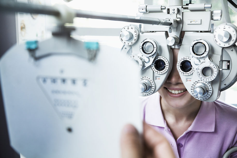
Non‑contact autorefractometer/autokeratometer Nidek ARK‑510A (Japan).

Computerized Refractometry and Keratometry

Computerised Tonometry
Non‑contact assessment of intra‑ocular pressure for comfortable diagnostics.
Measurement without corneal contact.
Ultra‑soft air pulse maximises patient comfort.
On‑screen display of all settings eliminates user error.
State‑of‑the‑art technology ensures accuracy and reliability.
Results correlate with Goldmann applanation tonometry standards.
Equipment: Leica/Reichert (USA‑Switzerland), Nidek Tono/Pachymeter‑Tonopachy NT‑530P (Japan).
Visual‑field examination in various ocular and neurological disorders.
Indicated for glaucoma, retinal pathology, optic‑nerve disease and neuro‑ophthalmic conditions.
Measures retinal contrast and threshold sensitivity.
Comparative software enables monitoring of disease progression.
Equipment: Centerfield Oculus Optikgeräte GmbH (Austria‑Germany).

Digital Computerised Perimetry

Ultrasonography
A‑B scanning for ocular evaluation and disease diagnosis.
A‑scan: measures anterior‑chamber depth, lens and ocular‑coat thickness, and axial length; performs IOL power calculation according to international formulas.
B‑scan: two‑dimensional imaging for vitreous assessment, documentation of retinal and choroidal detachment extent, detection and localisation of ocular and retro‑bulbar tumours, and foreign bodies.
Equipment: Alcon Ultrascan/Ultrasonik (USA‑Switzerland); Compact Touch Quantel Medical (France) – A‑B ultrasound scanner and pachymeter.

A/B ultrasound scanner, pachymeter Compact Touch, Quantel Medical
Provides a wide measurement range and easy data import. Applications – pachymetry, biometric measurements, A/B scanning (sectoral ultrasound with 2D image creation).


Computerised Topography and Ultrasonic Pachymetry
Accurate measurements prior to refractive surgery.
SIRIUS CSO, Italy – Before undergoing vision correction surgery, all patients undergo an ultrasound-based corneal thickness measurement. This method makes it possible to calculate the maximum permissible depth of treatment, which—depending on whether the case involves myopia, astigmatism, or hyperopia—determines how fully and safely the correction can be performed. The ultrasound device measures corneal thickness at 17 distinct points with an accuracy of up to 1 micron.
Ultrasonic corneal‑thickness measurement at 17 points with 1 μm resolution.
Pachymeter programmable to the physician’s parameters: measurement sequence, speed of sound (1000–2000 m/s), offset factor shape and value.
Probe: 20 MHz; tip diameter 1.2 mm; 45° angle.
Results stored in device memory and printable.
Equipment: SIRIUS CSO (Italy).


Clinic consultation appointment form
bottom of page